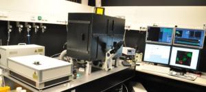
Olympus LSM FV1000
LSM unit: FluoView 1000
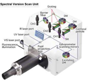
- Scanning method: two galvanometer scanning mirrors
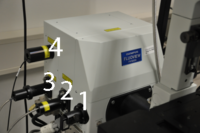
- 3 excitation light ports:
- 1: HBO lamp
- 2: Olympus laser box
- 3: Picoquant laser box
 Additional output port (4) connected to Avalanche photo diode detectors from Picoquant
- UV-Vis-IR Excitation with dichroic mirror turret, 6 positions (5 DMs plus 20/80 half mirror)
- Detection via 3 photomultiplier tubes (R7862, Hamamatsu, high sensitivity type)
- Ch1 and Ch2 equipped with 6 position beamsplitter turret and independent grating for fast and flexible spectral detection
- Specifications of grating units
- Resolution: < 2 nm
- Switching speed: 1 ms/100 nm
- Wavelength width: 1-100 nm
- Ch3 equipped with 6 position barrier filter turret
- The excitation laser can be imaged on another photomultiplier tube (R7400, Hamamatsu) to obtain transmission light images (see transmission channel ChT in paragraph Filter
- Scanning modes: X, Y, Z, t and λ, uni- or bidirectional scanning
- Image resolution: 64 x 64 - 4096 x 4096
- Fully motorized pinhole: displaceable in x and y-direction; diameter: 50 - 300 µm