Yokogawa CQ1
Overview
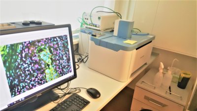
The CQ1 from Yokogawa is a benchtop high-content analysis system for quantitative image cytometry using spinning disk microscopy. CQ1 enables 3D imaging and quantification of live cell clusters, such as spheroids within a 3D culture vessel. Designed to quantitatively measure biological information from image data, it enables acquisition of information such as cell functions, signal transduction, cell mobility (such as invasion) or morphological information after automated image processing, which is rather difficult to obtain by conventional flow cytometric analysis.
The system contains 4 laser lines for 4-color fluorescence microscopy, a white LED for transmission bright field imaging and a fully motorized sample holder for long term automated aquisition. Axial drifts are compensated by a hardware autofocus device using a 785 nm laser. Detailed specifications can be found here or in the manual: CQ1_User_Manual.pdf.
The system contains 4 laser lines for 4-color fluorescence microscopy, a white LED for transmission bright field imaging and a fully motorized sample holder for long term automated aquisition. Axial drifts are compensated by a hardware autofocus device using a 785 nm laser. Detailed specifications can be found here or in the manual: CQ1_User_Manual.pdf.